153-2012-0258
来源:VET小柿子
感谢兽医英语口语班小伙伴曾杨茹的辛苦翻译。网站“兽医英语学习中心 (www.vetlearningcenter.cn)”上收录了我们班级翻译的资料,现在已经有大量X光片病例,皮肤病例,细胞学病例等等,点击文末阅读原文可以进入网站。口语班持续招生中,公众号后台回复“口语班”,了解详情。
来自Giuseppe Rubini
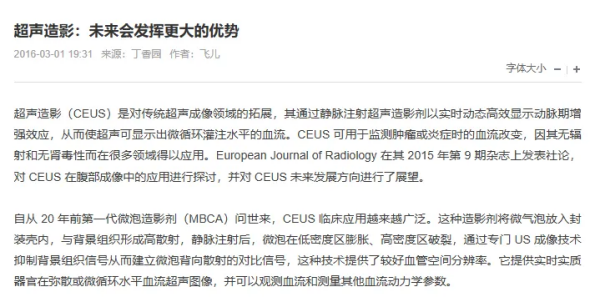
One year old female large breed crossbreed dog with mass growth in the last month. Cytology performed two weeks ago with hematoma. On ultrasound, see video, it appears to pertain to the abdominal muscles.
这只一岁雌性大型混种犬的肿块在过去1个月快速生长。2周前的细胞学检查提示血肿。超声结果如视频所示,这个肿块似乎与腹部肌肉相连。
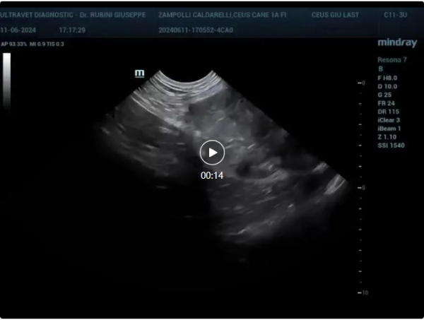
接下来是超声造影后的视频
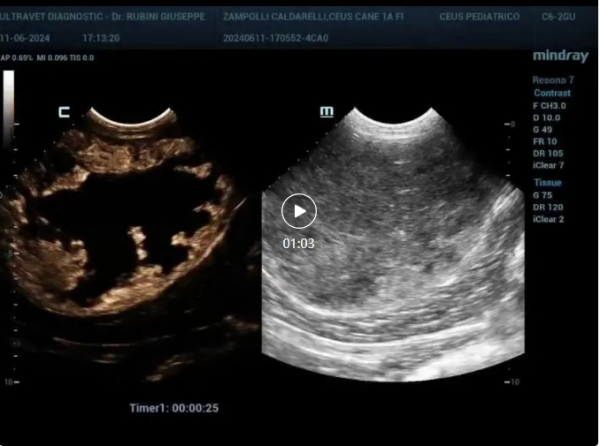
超声造影后发现肿块外周血管化。
这里就不详细讲血管结构了,很难用几句话解释。
这个超声造影结果提示恶性肿瘤,虽然年龄很小,但第一个怀疑的是血管肉瘤。
针对基础超声未检测到的血管化区域做了一个单独的细胞学,结果显示肌肉血管肉瘤。
正在等待CT分期结果、肿瘤学检查以及可能的组织学。
补充: